Scientists at Johns Hopkins University have cracked the code on how we develop sharp central vision before birth, pinpointing a crucial interplay between a vitamin A-derived molecule and thyroid hormones in the retina. This groundbreaking finding challenges a long-held theory about how essential light-sensing cells form and could pave the way for new treatments for vision-damaging diseases like macular degeneration and glaucoma.
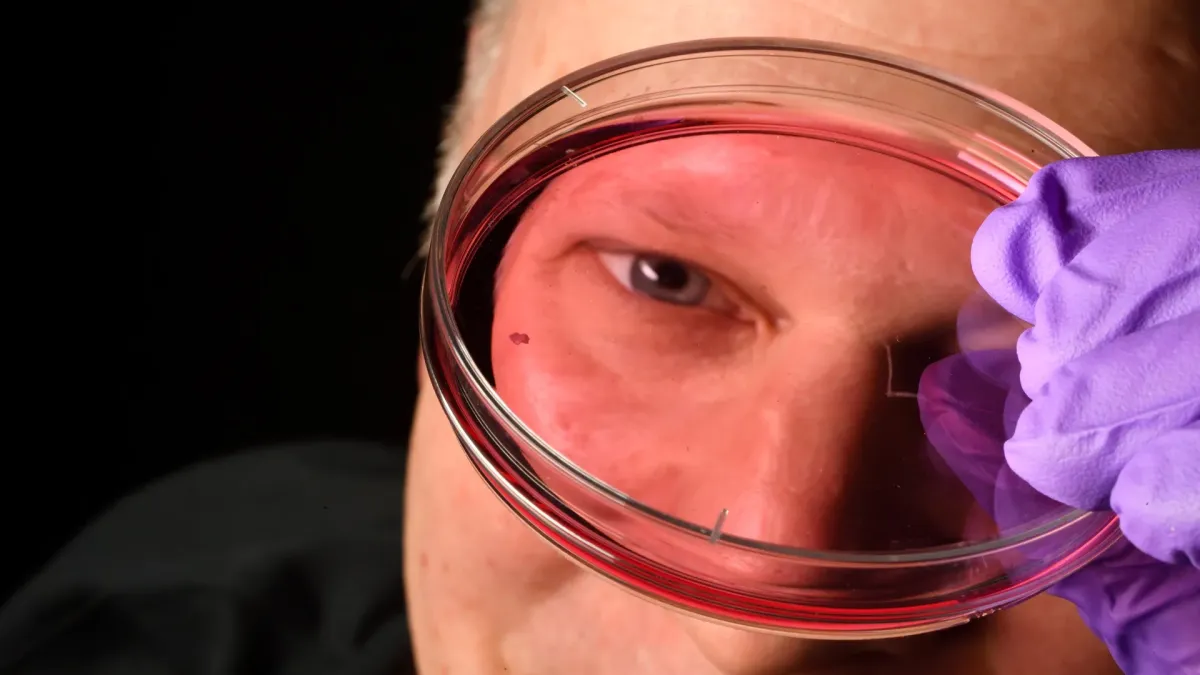
The research, conducted using lab-grown retinal tissue, revealed the intricate cellular events that shape the foveola – the tiny, bullseye-like region in the retina responsible for our sharpest vision. "This is a key step toward understanding the inner workings of the center of the retina," explained lead researcher Robert J. Johnston Jr., an associate professor of biology at Johns Hopkins. "By better understanding this region and developing organoids that mimic its function, we hope to one day grow and transplant these tissues to restore vision."
The team focused on cone photoreceptors, the cells that enable our daytime and color vision. While the foveola constitutes a small part of the retina, it accounts for roughly half of our visual perception and, surprisingly, only contains red and green cones, unlike the rest of the retina which has all three types (blue, green, and red). For decades, how this specialized pattern emerged remained a puzzle, partly because common lab animals don't share the same photoreceptor arrangement.
The new findings suggest a surprising transformation happens early in fetal development. Between weeks 10 and 12, a few blue cones appear in the developing foveola. By week 14, these cells transform into red and green cones. Researchers discovered this switch occurs via two main actions: first, retinoic acid (derived from vitamin A) reduces the creation of new blue cones, and then thyroid hormones encourage the remaining blue cones to convert into red and green ones.
"The main model in the field from about 30 years ago was that somehow the few blue cones you get in that region just move out of the way," Johnston noted. "Our data supports a different model. These cells actually convert over time, which is really surprising."
These discoveries could significantly impact future vision restoration efforts. Johnston's team is refining their retinal organoids to better replicate human retinal function, aiming to produce healthier photoreceptor cells for therapies that could potentially treat conditions like macular degeneration, which currently has no cure. "The goal with using this organoid tech is to eventually make an almost made-to-order population of photoreceptors," said study co-author Hussey. "A big avenue of potential is cell replacement therapy to introduce healthy cells that can reintegrate into the eye and potentially restore that lost vision."